


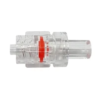


Flow rate is a critical parameter in CT imaging with contrast tubing, significantly influencing the quality of the images obtained and the overall diagnostic accuracy. As a leading supplier of CT Contrast Tubing, I have witnessed firsthand the importance of understanding how flow rate impacts CT imaging. In this blog, I will delve into the relationship between flow rate and CT imaging with contrast tubing, exploring the mechanisms at play and the practical implications for healthcare providers.
Understanding Contrast Media Delivery in CT Imaging
Before we discuss the impact of flow rate, it is essential to understand the role of contrast media in CT imaging. Contrast media are substances that are injected into the body to enhance the visibility of certain tissues and blood vessels during a CT scan. By increasing the contrast between different structures, contrast media allow radiologists to detect and diagnose a wide range of conditions more accurately.
Contrast media are typically delivered through a CT Contrast Tubing, which is connected to an intravenous (IV) catheter inserted into a patient's vein. The tubing is designed to withstand the high pressures generated by the contrast injection system and to ensure a smooth and consistent flow of contrast media into the patient's bloodstream.
The Role of Flow Rate in CT Imaging
Flow rate refers to the volume of contrast media that is injected into the patient's bloodstream per unit of time, typically measured in milliliters per second (mL/s). The flow rate plays a crucial role in determining the quality of the CT images obtained, as it affects the distribution and concentration of the contrast media in the body.
Image Enhancement
One of the primary goals of contrast-enhanced CT imaging is to maximize the contrast between the target tissue or blood vessel and the surrounding structures. A higher flow rate can help achieve this by rapidly delivering a large volume of contrast media to the area of interest, resulting in a higher concentration of contrast media in the target tissue and a more pronounced enhancement on the CT images.
For example, in a CT angiogram, which is used to visualize the blood vessels, a high flow rate is often required to ensure that the contrast media fills the vessels quickly and completely. This allows for clear visualization of the vessel lumen and the detection of any abnormalities, such as blockages or aneurysms.
Temporal Resolution
Another important aspect of CT imaging is temporal resolution, which refers to the ability to capture images at specific points in time. The flow rate of the contrast media affects the timing of the contrast enhancement in the body, and therefore, the temporal resolution of the CT scan.
A higher flow rate can result in a shorter transit time of the contrast media through the body, allowing for earlier and more precise imaging of the target tissue or blood vessel. This is particularly important in dynamic CT imaging, where sequential images are acquired over a period of time to observe the changes in contrast enhancement.
Patient Safety
While a higher flow rate can improve the quality of the CT images, it also increases the risk of adverse effects, such as contrast extravasation and nephrotoxicity. Contrast extravasation occurs when the contrast media leaks out of the blood vessel and into the surrounding tissue, causing swelling, pain, and potential tissue damage. Nephrotoxicity refers to the damage to the kidneys caused by the contrast media, which can lead to acute kidney injury.
Therefore, it is essential to balance the need for a high flow rate to achieve optimal image quality with the need to ensure patient safety. Healthcare providers must carefully consider the patient's age, weight, medical history, and renal function when determining the appropriate flow rate for a CT scan.
Factors Affecting Flow Rate
Several factors can affect the flow rate of the contrast media during a CT scan, including the size and length of the CT Contrast Tubing, the gauge of the IV catheter, the type of contrast injection system used, and the patient's blood vessel characteristics.
Tubing Size and Length
The size and length of the CT Contrast Tubing can have a significant impact on the flow rate. A larger diameter tubing allows for a higher flow rate, as it offers less resistance to the flow of contrast media. Similarly, a shorter tubing length can reduce the resistance and improve the flow rate.
However, it is important to note that using a larger diameter or shorter tubing may not always be feasible, as it may depend on the specific requirements of the CT scan and the patient's anatomy. Healthcare providers must carefully select the appropriate tubing size and length based on the patient's needs and the capabilities of the contrast injection system.
IV Catheter Gauge
The gauge of the IV catheter refers to the diameter of the catheter lumen, with a smaller gauge number indicating a larger diameter. A larger gauge catheter allows for a higher flow rate, as it offers less resistance to the flow of contrast media.
However, using a larger gauge catheter may not always be possible, especially in patients with small or fragile veins. In these cases, a smaller gauge catheter may be used, which may require a lower flow rate to ensure a safe and effective injection.
Contrast Injection System
The type of contrast injection system used can also affect the flow rate. There are two main types of contrast injection systems: power injectors and manual injectors.
Power injectors are capable of delivering contrast media at high flow rates and pressures, making them suitable for most contrast-enhanced CT scans. These injectors are typically programmable, allowing healthcare providers to adjust the flow rate, injection duration, and total volume of contrast media based on the patient's needs.


Manual injectors, on the other hand, rely on the operator's hand strength to inject the contrast media. While manual injectors are less expensive and more portable than power injectors, they are limited in terms of the flow rate and pressure they can generate. Therefore, manual injectors are typically used for lower flow rate applications or in situations where a power injector is not available.
Patient Factors
The patient's blood vessel characteristics, such as the size, depth, and elasticity of the veins, can also affect the flow rate. In patients with small or tortuous veins, it may be more difficult to achieve a high flow rate, as the veins may offer more resistance to the flow of contrast media.
In addition, the patient's cardiac output, which refers to the volume of blood pumped by the heart per minute, can also influence the flow rate. A higher cardiac output can result in a faster circulation of the contrast media in the body, allowing for a higher flow rate to be used.
Choosing the Appropriate Flow Rate
Choosing the appropriate flow rate for a CT scan requires careful consideration of several factors, including the type of CT scan being performed, the patient's age, weight, medical history, and renal function, and the capabilities of the contrast injection system.
In general, a higher flow rate is recommended for CT scans that require rapid and extensive contrast enhancement, such as CT angiograms and dynamic CT imaging. However, the flow rate should be adjusted based on the patient's individual needs and the specific requirements of the scan.
Healthcare providers should also consider the potential risks and benefits of using a higher flow rate, and take appropriate measures to minimize the risk of adverse effects. This may include using a smaller gauge catheter, reducing the injection pressure, or monitoring the patient closely during the scan.
Conclusion
Flow rate is a critical parameter in CT imaging with contrast tubing, as it affects the quality of the CT images obtained and the overall diagnostic accuracy. A higher flow rate can help achieve better image enhancement and temporal resolution, but it also increases the risk of adverse effects. Therefore, it is essential to carefully select the appropriate flow rate based on the patient's individual needs and the specific requirements of the scan.
As a supplier of CT Contrast Tubing, we understand the importance of providing high-quality tubing that can withstand the high pressures generated by the contrast injection system and ensure a smooth and consistent flow of contrast media into the patient's bloodstream. Our tubing is designed to meet the strictest quality standards and is available in a variety of sizes and configurations to meet the needs of different healthcare providers and patients.
If you are interested in learning more about our CT Contrast Tubing or other infusion therapy products, please contact us to discuss your specific requirements and to explore how we can help you achieve optimal results in your contrast-enhanced CT imaging procedures.
References
- Bushberg, J. T., Seibert, J. A., Leidholdt, E. M., & Boone, J. M. (2012). The essential physics of medical imaging. Lippincott Williams & Wilkins.
- McNitt-Gray, M. F. (2002). Physical principles of x-ray CT. Radiologic Clinics of North America, 40(2), 255-273.
- Samei, E., & Flynn, M. J. (2001). Image quality assessment: the use of phantoms in medical imaging. Physics in Medicine and Biology, 46(11), R153-R202.